【双语病例】肺泡蛋白沉积症PAP一例CT
发布时间:2022-10-22
发布时间:2022-10-22


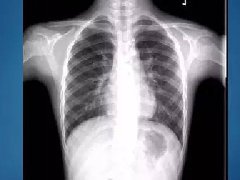
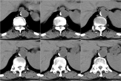
Findings
Differential diagnosis
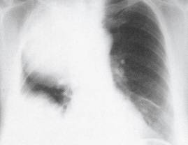
Diagnosis: Pulmonary alveolar proteinosis
The patient underwent bronchoalveolar lavage, which demonstrated milky and turbid fluid with thick sediment. Cytology demonstrated large foamy macrophages. Serum antigranulocyte-macrophage colony-stimulating factor (anti-GM-CSF) autoantibodies were positive.

上一篇:胸腺囊肿
下一篇:纵隔支气管囊肿1例CT影像表现